EMERGENCY MEDICINE GUIDEWIRE
  • HOME
  • The Podcasts
    • Core Concepts
    • Critical Care Topics
    • Intern Nuggets
    • Pediatric EM
    • Toxicology
    • Trauma Topics
    • Sports Medicine Corner
  • Cards for EM
    • ECG Monthly Lesson
  • CMC Imaging Mastery Project
    • Adult Chest X-Ray Cases
    • Pediatric X-Ray Cases
    • Pediatric Orthopedic X-Ray Cases
    • Abdominal Imaging Room
    • Implanted Device Radiology Room
    • Condition Specific Radiology
  • Global EM
  • Shownotes
    • Core Concepts ShowNotes
    • Cards for EM ShowNotes
    • Ped EM Morsels ReBaked Shownotes
    • Pediatric EM Shownotes
    • Sports Med Shownotes
    • Toxicology Shownotes
    • Trauma Talks Shownotes
  • Subscribe
    • iTunes
    • Google Play
    • Stitcher
  • About
  • Associated Sites
    • CMCEdMasters
    • CMCECGMasters
    • PedEMMorsels
Search by typing & pressing enter

YOUR CART

Dr. Littmann's Monthly ECG Lessons

9/27/2020 0 Comments

September 2020 ECG Lesson: Wellens Syndrome

By: Quinton Nannet, MD and Dominic Nicacio, MD
​Editors: Drs. Littmann and Gibbs 
Picture
Quinton Nannet, MD
Picture
Dominic Nicacio, MD
Case:
A 56-year-old man with a medical history of schizophrenia, tobacco abuse, and coronary artery disease presented to our emergency department for chest pain. The patient had drug-eluting stents placed to the mid-circumflex and right coronary arteries 13 months before the current presentation. He reports that he has not been taking aspirin or clopidogrel due to financial limitations. He also continues tobacco use of ½ pack a day. The patient describes 3 weeks of severe left-sided, exertional chest pain and associated dyspnea. The pain is normally relieved with rest and ibuprofen, but over the past two days he has had similar pain at rest with only intermittent relief. 
 
The following electrocardiogram was obtained:
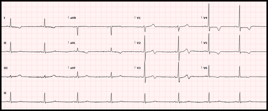
Picture
ECG Interpretation:
  • Rate: 48/min
  • Rhythm: Sinus bradycardia
  • Axis: Normal QRS axis
  • QRS: <120 msec; no abnormal Q waves
  • ST segments: No ST elevation or ST depression
  • T waves: Inverted T waves in I, aVL, V2-V6 with biphasic (positive-negative) T waves in V2 and V3 classified as Wellens syndrome type A
Diagnosis:
Wellens syndrome
Significance of Findings:
  • Wellens syndrome is a pattern of electrocardiographic T-wave changes associated with usually severe or critical proximal left anterior descending (LAD) coronary artery stenosis. The syndrome is also referred to as LAD coronary T-wave syndrome.
  • The T-wave abnormalities are seen in the anterior chest leads, usually from V1-V4
  • The T-wave changes are persistent and may remain in place for hours to weeks; making it likely that the clinician will encounter these changes when the patient is chest pain-free.
  • Pathophysiology: not clearly established. According to one theory, the LAD experiences an occlusion that would result in a STEMI if captured on the ECG. This occlusion is reperfused either from spontaneous lysis, from aspirin, or resolution of vasospasm leading to pain relief and progressing to biphasic T waves and then deeply inverted T waves. Since the artery is highly unstable, a recurrent occlusion may progress to tall “hyperacute” T waves or frank ST elevation myocardial infarction (STEMI).
  • Wellens type A ECG pattern
    • T wave morphology: biphasic, initially positive and terminally negative
    • Prevalence: ~ 25% of Wellens syndrome patients
    • Significance: represents an early phase of Wellens syndrome with T wave morphology then progressing toward type B T waves
  • Wellens type B ECG pattern
    • T wave morphology: symmetric and deeply inverted
    • Prevalence: ~ 75% of Wellens syndrome patients
    • Significance: represents a later phase of Wellens syndrome
  • The same ECG may show both type A and type B patterns in different chest leads
  • The ECG findings may persist for weeks:
Picture
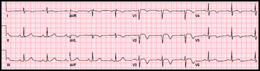
Image Source: https://www.ncbi.nlm.nih.gov/books/NBK482490/
 
Diagnostic criteria of Wellens syndrome:
  • Presentation with acute, subacute, recent or stuttering chest pain (probable acute coronary syndrome)
  • Biphasic (positive-negative) or deeply inverted, usually symmetrical T waves in leads V1-V4
  • Isoelectric or minimally elevated (<1 mm) ST segments
  • Preservation of precordial R-wave progression and no abnormal Q waves
  • ECG pattern may be present in a pain-free state
  • Normal or slightly elevated cardiac biomarkers
Management:
  • Immediate cardiac consultation is warranted
  • Patients should be treated with aspirin and heparin
  • Definitive management is heart catheterization with LAD stent placement
  • In Wellens syndrome, early revascularization is far superior to medical management
  • Without appropriate intervention, patients are at high risk for STEMI with significant morbidity and mortality
Patient Disposition:
The patient was admitted to the interventional cardiology service. Cardiac catheterization revealed 99% stenosis of the left main coronary artery, 50% stenosis of the left anterior descending coronary artery and 100% occlusion of the previously stented left circumflex. The previously stented right coronary artery was patent.

Results of cardiac catheterization are shown below:

Picture
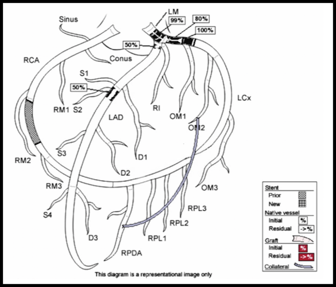
Interventions:
Due to the extent of stenoses, the patient had an intra-aortic balloon pump placed to reduce myocardial oxygen demand. The next day, he underwent multivessel coronary artery bypass grafting.
 
Socio-economic considerations:
This case highlights the importance of dual antiplatelet therapy and smoking cessation in patients with stent placement. It also demonstrates the challenges mental health and socioeconomic factors play in the patients’ ability to adhere to treatment plans.
Dr. Littmann’s comments:
There are 4 major types of high-risk acute coronary syndrome (ACS) where the ECG does not show diagnostic ST-segment elevation. These include:
  • Posterolateral MI (old nomenclature: true posterior or high posterior MI). This diagnosis should be suspected in patients who present with typical ACS and the ECG shows ST depression in the anterior chest leads. The diagnosis can be confirmed by placing ECG leads to the back. This condition is a STEMI equivalent that warrants emergent cardiac catheterization and reperfusion.
  • De Winter sign. This diagnosis should be suspected in patients who present with typical ACS and the ECG shows depression of the ST segment at the J point followed by tall, symmetrical upright T waves (“hyperacute T waves”). It usually signifies total or subtotal acute occlusion of the LAD. In my view, this too should be considered a STEMI equivalent and patients should undergo emergent cardiac catheterization and reperfusion. See the August 2020 ECG blog for details.
  • aVR sign. This is characterized by acute chest pain, diffuse ST-segment elevation and ST elevation in aVR of ³ 1 mm. The aVR sign is strongly suggestive of severe and diffuse ischemia due to tight left main or multivessel coronary artery disease. However, it is not a STEMI equivalent. If other important causes such as proximal aortic dissection and hemorrhagic shock have been ruled out, the patients should undergo relatively urgent cardiac catheterization.
  • Wellens syndrome. This is characterized by acute, subacute or stuttering chest pain, biphasic (positive-negative) and/or deep symmetrical T waves in the anterior chest leads and normal or slightly elevated troponins. The patients may be chest pain free on presentation.
    • Of the above listed 4 conditions, the Wellens syndrome is the “oldest” first described in 1982 and therefore, it is the best known among emergency medicine providers.
    • In my view, however, of the above listed conditions it is the least worrisome that usually does not require emergent intervention. In the original description of the Wellens syndrome, those patients who presented with ACS with the Wellens sign in the ECG and did not undergo bypass surgery (there was no PCI available at that time), had a significant likelihood of progressing to STEMI in the weeks or months after presentation. Also, medical management in the 1970s-1980s only included beta blocker and nitrates; we did not even use aspirin, heparin or statin.
    • In general, patients with ACS, ongoing chest pain and/or elevated troponin routinely undergo cardiac catheterization regardless of Wellens or not Wellens.
    • Those patients, however, who are pain free and have negative troponin and no Wellens sign usually first undergo noninvasive evaluation and then cardiac catheterization if the noninvasive test shows high-risk abnormalities.
    • In patients with ACS and the Wellens-type T-wave abnormalities, however, noninvasive testing should usually be avoided. Most patients should undergo cardiac catheterization even if they are pain free and have negative troponins.
    • Wellens-type T-wave changes are not completely specific for tight proximal LAD stenosis. They can be present in patients with massive pulmonary embolism, hypertensive urgency, acute or acute-on-chronic renal failure and severe hypertension. In questionable cases, adenosine Myoview, Lexiscan Myoview or CT coronary angiogram can be safely performed. Frank exercise testing and dobutamine studies, however, should not be done because they can provoke acute MI.​
References:
  • doi: 10.1053/ajem.2002.34800
  • Miner B, Grigg WS, Hart EH. Wellens Syndrome. [Updated 2020 May 28]. In: StatPearls [Internet]. Treasure Island (FL): StatPearls Publishing; 2020 Jan-. Available from: https://www.ncbi.nlm.nih.gov/books/NBK482490/
  • doi: 10.1161/CIRCULATIONAHA.119.043780
0 Comments

    Author

    This blog represents important ECG lessons that the Emergency Medicine Residents from Carolinas Medical Center (Charlotte, NC) rotating through the Cardiology service encounter.  Test your knowledge with them! The esteemed educators Dr. Laszlo Littmann and Dr. Michael Gibbs serve as the primary content editors and course directors. 

    Archives

    September 2020
    August 2020
    July 2020
    May 2020
    March 2020
    January 2020
    October 2019
    August 2019

    Categories

    All Bradycardia Cardiology ECG Lesson Heart Block Syncope Ventricular Tachycardia Vtach

    RSS Feed

EMGuideWire

Picture
From the J. Lee Garvey Innovation Studio in the 
Department of Emergency Medicine
Carolinas Medical Center
Charlotte, NC

Picture
© COPYRIGHT 2015. ALL RIGHTS RESERVED.
LEGAL DISCLAIMER (to make sure that we are all clear about this):The information on this website and podcasts are the opinions of the authors solely.

For Health Care Practitioners: This website and its associated products are provided only for medical education purposes. Although the editors have made every effort to provide the most up-to-date evidence-based medical information, this writing should not necessarily be considered the standard of care and may not reflect individual practices in other geographic locations.

​For the Public
: This website and its associated products are not intended to be a substitute for professional medical advice, diagnosis, or treatment. Your physician or other qualified health care provider should be contacted with any questions you may have regarding a medical condition. Do not disregard professional medical advice or delay seeking it based on information from this writing. Relying on information provided in this website and podcast is done at your own risk. In the event of a medical emergency, contact your physician or call 9-1-1 immediately.