EMERGENCY MEDICINE GUIDEWIRE
  • HOME
  • The Podcasts
    • Core Concepts
    • Critical Care Topics
    • Intern Nuggets
    • Pediatric EM
    • Toxicology
    • Trauma Topics
    • Sports Medicine Corner
  • Cards for EM
    • ECG Monthly Lesson
  • CMC Imaging Mastery Project
    • Adult Chest X-Ray Cases
    • Pediatric X-Ray Cases
    • Pediatric Orthopedic X-Ray Cases
    • Abdominal Imaging Room
    • Implanted Device Radiology Room
    • Condition Specific Radiology
  • Global EM
  • Shownotes
    • Core Concepts ShowNotes
    • Cards for EM ShowNotes
    • Ped EM Morsels ReBaked Shownotes
    • Pediatric EM Shownotes
    • Sports Med Shownotes
    • Toxicology Shownotes
    • Trauma Talks Shownotes
  • Subscribe
    • iTunes
    • Google Play
    • Stitcher
  • About
  • Associated Sites
    • CMCEdMasters
    • CMCECGMasters
    • PedEMMorsels
Search by typing & pressing enter

YOUR CART

Dr. Littmann's Monthly ECG Lessons

8/27/2020 0 Comments

August 2020 ECG Lesson: "That is an Interesting Sign"

BY: ERIC SABATINI-REGUEIRA, MD and COURTNEY FLEMING, MD
EDITORS: DRS. LITTMANN and GIBBS
Picture
Eric Sabatini-Regueira, MD
Picture
Courtney Fleming, MD
Case:
A 36-yo obese female with no other known past medical condition presented to the emergency department with a 30-minute history of chest pain. The pain was described as being a retrosternal pressure-like discomfort that was non-radiating, and 10/10 in severity. The patient denied having similar episodes in the past. There were no alleviating or worsening factors. During the physical examination, the patient was noted to have the Levine’s sign (clenched fist held over her chest).
 
The following electrocardiogram was obtained:
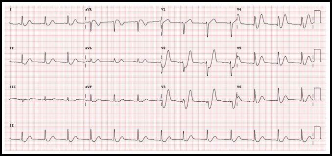
Picture
ECG Interpretation
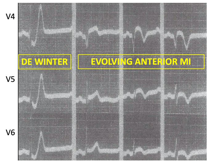
Rate: 78/min
Rhythm: Normal sinus rhythm
Axis: Normal QRS axis
QRS: <120 msec; no abnormal Q waves
ST-T: ST depression at the J point (at the junction between the QRS complexes and ST segments) in the anterior/lateral leads, mostly upsloping ST segments and then, very tall peaked T waves (de Winter T waves). ST segment elevation in aVR.
Significance of ECG Finding

  • Thought to be first described by Dr. Robbert Jan de Winter in 2008 in his letter to the editor to the New England Journal of Medicine, the pattern of peaked T-waves as a sign of myocardial ischemia was actually first noted by Dr. William Dressler in 1947 in a case series of over 27 patients. What de Winter defined was “1- to 3-mm upsloping ST-segment depression at the J point in leads V1 to V6 that continued into tall, positive symmetrical T waves” that are often accompanied by a “1- to 2-mm ST-elevation in lead aVR”.
  • The de Winter pattern is significant because it is associated with occlusion of the proximal left anterior descending coronary artery (LAD) if found in the ECG of patients with chest pain or a history suspicious for acute coronary syndrome. In fact, this patytern is found in 2% of patients diagnosed with proximal occlusion of the LAD, with an associated positive predictive value of 95-100%. The other 98% of patients with acute LAD occlusion had frank STEMI in the ECG. As such, it is critical not to overlook the de Winter pattern which is not common, but is associated with important clinical outcomes.
  • The pattern has been found to be more common in young males (mean age 52 compared with mean age of 61 in patients with STEMI on ECG) and in ptients with history of hypercholesterolemia, smoking and family history of coronary artery disease.
  • The exact pathophysiologic mechanism of why the de Winter pattern occurs in patients with occlusion of the proximal LAD is largely unknown. It has been speculated that it may be due to endocardial conduction delay due to anatomical variations in the Purkinje fibers. Other theories attribute the pattern to variations in coronary anatomy and collateral recruitment.
  • There has been much debate in the medical literature in regard to whether the de Winter pattern should be considered a STEMI equivalent. Many studies have defined the de Winter pattern as being a static sign that occurs very early after symptom onset and persists until reperfusion therapy. However, recent studies have identified the de Winter pattern as a more dynamic entity that can either evolve into classic ST-elevation or occur after the resolution of ST-elevation on the ECG. The theory behind this is that the de Winter pattern represents subtotal occlusion of the LAD, and progression to frank STEMI occurs with continued thrombus formation towards total arterial occlusion. Conversely, resolution from STEMI to a de Winter pattern could represent autolysis of a totally occlusive thrombus back to subtotal occlusion. Regardless if the pattern is static or dynamic, it does remain highly suggestive of a high degree of arterial occlusion.
  • There are few definitive guidelines for management of patients who present with symptoms suggestive of ACS and the de Winter pattern on the ECG. Most recent guidelines by the American Heart Association do not explicitly recommend percutaneous intervention or outline thrombolysis management for patients presenting with the de Winter sign as a singular indication for acute coronary intervention. Though rare, specific guidelines for management of patients with de Winter pattern is warranted as studies show this important ECG sign is often missed by clinicians, and delay in diagnosis can lead to higher total ischemic time and possible higher mortality. In the emergency department, this should include immediate cardiology consultation and when available, immediate bedside echocardiography to assess for the presence of left ventricular wall motion abnormalities.
Disposition
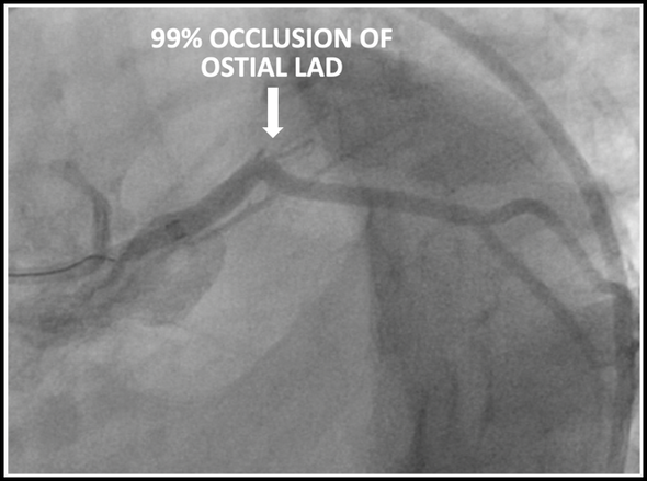
The patient underwent emergent cardiac catheterization which found a 99% occlusion of the ostial LAD. The initial troponin-T was 4.78ng/mL and peaked at >50ng/mL (detection limit) later that day. It is important to remember that a positive troponin is not needed to make the diagnosis of STEMI or a STEMI equivalent.

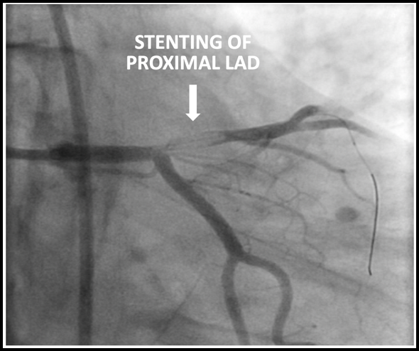
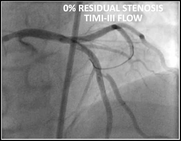
Results of cardiac catheterization and stenting of the proximal LAD are shown below
Picture
Picture
Picture
Dr. Littmann’s comments:
Drs. Sabatini-Regueria and Fleming have beautifully summarized our current understanding of the de Winter sign, its prevalence, clinical significance, possible mechanism and the somewhat controversial guideline recommendations how to proceed when it is manifest. I have a few comments about the de Winter sign:
  • The original description of the de Winter sign requires depression of the ST segment at the J point followed by very tall T waves. In my view, the ST depression part is not mandatory; very tall (“hyperacute”) T waves alone are equally diagnostic of proximal LAD occlusion (see example below).
  • Unfortunately, there are no absolute diagnostic criteria what constitute hyperacute T waves, how tall the T waves need be. The clinical scenario is most important: if a patient has acute chest pain suggestive of acute MI, very tall T waves in the chest leads should warrant emergent catheterization and reperfusion.
  • How can one distinguish tall T waves of hyperkalemia from tall T waves of de Winter? The clinical scenario is usually completely different. In a patient with sepsis, shock and renal failure, tall T waves almost certainly indicate hyperkalemia. In a patient who presents with acute chest pain, it is most likely the de Winter sign. Also, tall T waves of hyperkalemia are usually narrow based whereas the de Winter T waves are usually not.
  • Although it is uncertain why 98% of patients with acute occlusion of the proximal LAD present with frank STEMI and 2% with the de Winter sign, the most compelling evidence suggests that the de Winter sign signifies subtotal occlusion of the proximal LAD, and STEMI signifies total occlusion. That is probably why most de Winter presentations are very early after the onset of chest pain and why, if not reperfused, the de Winter sign usually progresses to frank STEMI.
  • In my view with a clinical presentation of acute chest pain, the de Winter sign should unequivocally be considered a STEMI equivalent mandating emergent cath and reperfusion. A lot is at stake!
  • What is now known as the de Winter sign was originally described in 1947 by Dr. Dressler, the same cardiologist who described the Dressler syndrome. Although the de Winter group deserves huge credit for presenting more details about the de Winter sign and for popularizing it, in my view it should be renamed the Dressler – de Winter sign.
  • Dr. de Winter is from the cardiology group in the Netherlands which used to be headed by Dr. Wellens. Both on the de Winter article and on the Wellens sign article, Dr. Wellens is the last author. They not only described these two syndromes, but also were one of the first to study the aVR sign. Dr. Wellens was a giant in electrocardiology and cardiac electrophysiology. He passed away a few months ago at age 85. The cardiology world is mourning his loss.
​
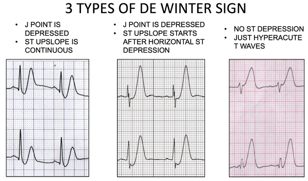
Below are the three types of de Winter ECGs. Note that in the third type, there is no ST depression, just hyperacute T waves.
Picture
Below is an edited version of the de Winter sign as described by Dr. Dressler in 1947
Picture
​References (all represented by DOI numbers):
10.1056/NEJMc0804737
10.1016/0002-8703(47)90343-8
10.1016/j.jacc.2018.08.1038
10.1016/j.ajem.2013.09.037
10.12998/wjcc.v7.i20.3296
10.4103/HEARTVIEWS.HEARTVIEWS_90_19
10.1155/2018/6868204
10.21037/atm.2019.07.19
10.1016/j.jelectrocard.2017.08.024
10.1016/j.jacc.2020.07.015
0 Comments

    Author

    This blog represents important ECG lessons that the Emergency Medicine Residents from Carolinas Medical Center (Charlotte, NC) rotating through the Cardiology service encounter.  Test your knowledge with them! The esteemed educators Dr. Laszlo Littmann and Dr. Michael Gibbs serve as the primary content editors and course directors. 

    Archives

    September 2020
    August 2020
    July 2020
    May 2020
    March 2020
    January 2020
    October 2019
    August 2019

    Categories

    All Bradycardia Cardiology ECG Lesson Heart Block Syncope Ventricular Tachycardia Vtach

    RSS Feed

EMGuideWire

Picture
From the J. Lee Garvey Innovation Studio in the 
Department of Emergency Medicine
Carolinas Medical Center
Charlotte, NC

Picture
© COPYRIGHT 2015. ALL RIGHTS RESERVED.
LEGAL DISCLAIMER (to make sure that we are all clear about this):The information on this website and podcasts are the opinions of the authors solely.

For Health Care Practitioners: This website and its associated products are provided only for medical education purposes. Although the editors have made every effort to provide the most up-to-date evidence-based medical information, this writing should not necessarily be considered the standard of care and may not reflect individual practices in other geographic locations.

​For the Public
: This website and its associated products are not intended to be a substitute for professional medical advice, diagnosis, or treatment. Your physician or other qualified health care provider should be contacted with any questions you may have regarding a medical condition. Do not disregard professional medical advice or delay seeking it based on information from this writing. Relying on information provided in this website and podcast is done at your own risk. In the event of a medical emergency, contact your physician or call 9-1-1 immediately.