|
11/2/2020 0 Comments Severe Asthma Management in the ED
Therapy Options:
0 Comments
9/9/2020 0 Comments Bradycardia Basics
2/25/2020 0 Comments Pregnancy and Trauma
OB Trauma
-Travis Barlock ---- See also Slides from Dr. Michael Gibbs on this subject - http://www.mededmasters.com/obgyn.html 1/15/2020 0 Comments SVC Syndrome
Topic: Superior Vena Cava Syndrome
Definition:
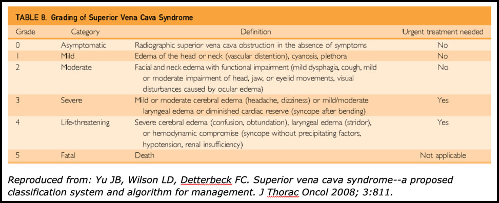
References:
7/30/2019 0 Comments Skin and Soft Tissue Infections
Skin and Soft Tissue Infections
Epidemiology
Important Aspects of your History
The Mimics
Workup
Treatment
“Special” cellulitis
Necrotizing fasciitis +/- Toxic Shock Syndrome
Republished with Permission from www.PedEMMorsels.com
Difficult Airway: Basics
Difficult Airway: Important Differences Anatomic and physiologic differences influence how the oral, pharyngeal, and tracheal axes align as well as how the respiratory mechanics function and how the child compensates to physiologic stress. The differences are most prominent in children < 2 years of age and more adult anatomy evolves as children progress to 8 years of age. Anatomic Differences:
Physiologic Differences:
Difficult Airway: Predictive Factors
Difficult Airway: Assume the Worst
Moral of the Morsel
Taking care of children in the ED isn't just diagnosing viruses - child abuse/neglect is more prevalent than we'd like to think:
- 2-10% of children presenting to the ED are victims of abuse or neglect - children that are abused often have multiple healthcare visits before it is recognized Child maltreatment includes neglect and abuse Neglect - failure to meet the most basic needs of a child (food, shelter, supervision, nurturance) - can be educational, psychological, emotional, medical neglect, or supervisory - by far the most common type of maltreatment - about ⅔ of cases - very difficult to identify diagnose Abuse - includes physical abuse, sexual abuse, emotional abuse, and medical child abuse * medical child abuse is the current term for Munchausen Syndrome by proxy - physical abuse (also referred to as non-accidental trauma or NAT) is often the most recognizable type of abuse - makes up 16% of maltreatment cases Providers should be mindful of sentinel injuries - injuries without a plausible explanation - knowing basic developmental milestones can be helpful 4-5 months - rolling over 6 months - sitting unassisted 9 months - pulling to a stand and walking 12 months - walking **soft tissue injuries - bruising in children who cannot cruise, or in high risk areas - normal childhood bruises occur on surfaces that take impact when the child falls bruising on the knees and shins isn’t alarming bruising on areas like the back, buttocks, thighs, abdomen is concerning - TEN-4 FACES P can help you identify injuries that suggest potential abuse * bruising on the Trunk, Ears, or Neck in a child less than 4 years old * ANYbruising on a child less than 4 months * injuries/bruising to the Frenulum, Auricular area, Cheek, Eyes, Sclera or * Patterned bruising is a red flag **skeletal injuries are the second most common presentation of abuse - certain fractures should raise your suspicion for abuse - rib fractures (make sure you check that CXR ordered to look for pneumonia) - any fracture in a child that cannot walk - long bone fractures in an infant or toddler **abusive head trauma (formally known as “shaken baby syndrome”) - this is the most common cause of death following abuse - 30% of cases are missed initially – remember to consider it in cases of excessive fussiness or altered mental status **chest and abdominal injuries - the abdomen can hide injuries and hold a lot of blood - you won’t always see bruising - elevated liver enzymes or lipase should raise your concern for occult intra-abdominal injury What can you do to help prevent a missed diagnosis of abuse?
ACEP Now. The Recognition of Child Abuse. May 1, 2012. Colbourne M, Clarke MS. Child Abuse and Neglect. In: Tintinalli JE, Stapczynski J, Ma O, Yealy DM, Meckler GD, Cline DM. eds. Tintinalli’s Emergency Medicine: A Comprehensive Study Guide, 8e New York, NY: McGraw-Hill; 2016. http://accessmedicine.mhmedical.com.libproxy.lib.unc.edu/content.aspx?bookid=1658§ionid=109435341. Accessed May 15, 2019. Christian CW. The Evaluation of Suspected Child Abuse. PEDIATRICS. 2015; 135 (5): e1337-e1354. Glick JC, Lorand MA, and Bilka KR. Physical Abuse of Children. Pediatrics in Review. 2016; 37(4): 146-158. Lindberg DM, Beaty B, Juarez-Colunga E, et al. Testing for Abuse in Children With Sentinel Injuries. PEDIATRICS. 2015; 136(5): 831-838. Petska HW and Sheets LK. Sentinel Injuries Subtle Findings of Physical Abuse. Pediatric Clinics of North America. 2014; 61(5): 923-935. Ravichandiran N, Schuh S, Bejuk M, et al. Delayed Identification of Pediatric Abuse-Related Fractures. PEDIATRICS. 2009; 125 (1): 60-66. Sheets LK, Leach ME, Koszewski IJ, et al. Sentinel Injuries in Infants Evaluate for Child Physical Abuse. PEDIATRICS. 2013; 131(4): 701-107. Teeuw AH, Derkx BHF, Koster WA, et al. Detection of child abuse and neglect in the emergency room. European Journal of Pediatrics. 2012; 171: 877-885. Teeuw AH, Hoytema van Konijnenburg EM, Sieswerda-Hoogendoorn T, et al. Parents’ Opinions About a Routine Head-to-Toe Examination of Children as a Screening Instrument for Child Abuse and Neglect in Children Visiting the Emergency Department. Journal of Emergency Nursing. 2016; 42(2): 128-138. 3/22/2019 0 Comments Myasthenic Crisis
MG Pathophysiology & Symptoms
MG Crisis (MC) Causes
Drugs to avoid in MG Patients:
Drugs that should be used with caution:
Evaluation
Focused 30-second Neuro Exam
Airway Management
- Vital capacity less than 10-20 mL/kg - Single-breath test (unable to count past 20) - Unable to clear secretions
Management After Stabilization
Sources: Bird, S.J. & Levine, J.M (2019). Myasthenic crisis. In A.F. Eicheler & G. Finlay (Ed.), UpToDate.Retrieved February 27, 2019, from https://www.uptodate.com/contents/myasthenic-crisis Howard, J.F. (2009). Myasthenia Gravis: A manual for the health care provider. Myasthenia Gravis Foundation of America, Inc. Jowkar, A. (2010). Neuromuscular topics for Neuroscience ICU RNs. Presentation, University of North Carolina at Chapel Hill. Roper, J., Fleming M.E., Long. B. et al. (2017). Myasthenia Gravis and Crisis: Evaluation and Management in the Emergency Department. The Journal of Emergency Medicine, 53(5): 843-853. http://dx.doi.org/10.1016/j.jemermed.2017.06.099 Tintinalli, J. E., Stapczynski, J. S., Ma, O. J. et al. (2016). Tintinalli's emergency medicine: A comprehensive study guide(Eighth edition.). New York: McGraw-Hill Education. |
 RSS Feed
LEGAL DISCLAIMER (to make sure that we are all clear about this):The information on this website and podcasts are the opinions of the authors solely.
For Health Care Practitioners: This website and its associated products are provided only for medical education purposes. Although the editors have made every effort to provide the most up-to-date evidence-based medical information, this writing should not necessarily be considered the standard of care and may not reflect individual practices in other geographic locations.
For the Public: This website and its associated products are not intended to be a substitute for professional medical advice, diagnosis, or treatment. Your physician or other qualified health care provider should be contacted with any questions you may have regarding a medical condition. Do not disregard professional medical advice or delay seeking it based on information from this writing. Relying on information provided in this website and podcast is done at your own risk. In the event of a medical emergency, contact your physician or call 9-1-1 immediately.