EMERGENCY MEDICINE GUIDEWIRE
  • HOME
  • The Podcasts
    • Core Concepts
    • Critical Care Topics
    • Intern Nuggets
    • Pediatric EM
    • Toxicology
    • Trauma Topics
    • Sports Medicine Corner
  • Cards for EM
    • ECG Monthly Lesson
  • CMC Imaging Mastery Project
    • Adult Chest X-Ray Cases
    • Pediatric X-Ray Cases
    • Pediatric Orthopedic X-Ray Cases
    • Abdominal Imaging Room
    • Implanted Device Radiology Room
    • Condition Specific Radiology
  • Global EM
  • Shownotes
    • Core Concepts ShowNotes
    • Cards for EM ShowNotes
    • Ped EM Morsels ReBaked Shownotes
    • Pediatric EM Shownotes
    • Sports Med Shownotes
    • Toxicology Shownotes
    • Trauma Talks Shownotes
  • Subscribe
    • iTunes
    • Google Play
    • Stitcher
  • About
  • Associated Sites
    • CMCEdMasters
    • CMCECGMasters
    • PedEMMorsels
Search by typing & pressing enter

YOUR CART

5/3/2019 0 Comments

Sudden Cardiac Death in Athletes


​Sudden cardiac death = most frequent medical cause of sudden death in athletes
  • A recent estimate of SCD incidence ranged from 1 in 40,000 to 1 in 80,000 athletes per year
 
Jason Collier, 28 (2005), basketball
Eddie Guerrero, 38 (2005), wrestling
Fab Melo, 26 (2017), basketball
 
What is an “athlete?”
  •  “One who participates in an organized team or individual sport that requires competition against others as a central component, places a high premium on excellence and achievement, and requires some form of systematic (and usually intense) training.” 
 
When estimating SCD incidence, the population of athletes “at risk” may be difficult to quantify
 
Definition? 
  1. Deaths with exertion or shortly (< 1 hour) after exertion, 
  2. Any SCD in an athlete (exertional or outside of exertion) and also episodes of resuscitated sudden cardiac arrest
 
These inconsistencies help account for the wide range of estimated incidence of SCD in athletes in prior reports, from 1 in 3,000 up to 1 in 1 million
 
Risk among male Division 1 basketball players = 10x that in the overall athlete population
  • Mechanism is NOT clear
  •  Marfan syndrome and resulting aortic dissection have most commonly occurred in male basketball players, this accounts for a small fraction of the deaths in this population
 
Causes
  • Athletes <35
    • Inherited cardiac conditions predominate: hypertrophic cardiomyopathy and anomalous origin of a coronary artery (two most common causes in the United States)
  • Athletes >35
    • Most SCD events are due to acquired atherosclerotic coronary artery disease 
  • Many of these diagnoses may not be clinically apparent and first symptom may be sudden death
  • Approximately 30% of athletes with SCD have been reported to have had symptoms such as chest pain, shortness of breath, performance decline, palpitations, near syncope, or syncope leading up to the event
 
The Young Athlete
  • MCC = HCM
    • Genetic condition characterized by left ventricular hypertrophy and cardiac myocyte disarray
      • Predisposes to ventricular arrhythmias
    • Prevalence = 1 in 200 individuals
    • First presenting symptom may be Sudden Cardiac Death (SCD)
  • Congenital coronary artery anomalies, consisting of a variety of abnormalities of coronary origin and proximal course, are the second most common cause of SCD
  • The remaining causes of SCD in athletes include other inherited or acquired myocardial diseases, other structural cardiovascular disease, or primary arrhythmogenic disorders
  • Recently, data from NCAA athletes suggests that the most common causes of SCD may merit re-evaluation.
    • Most common finding at autopsy for SCD cases was a structurally normal heart (25%), implying that arrhythmias and other electrical disorders may be the most common etiology.
    • Coronary anomalies were the second most common finding and were present at a similar proportion as was previously reported, but definitive HCM was far less frequent compared to prior information
  • Reports from other countries such as Italy and Denmark have found that the most common cause of SCD in young athletes is arrhythmogenic right ventricular cardiomyopathy (ARVC)-- the artist formerly known as ARVD
    • ARVC is a genetic cardiomyopathy that is typified by fibro-fatty infiltration and dilation of the right ventricle
    • Risk of ventricular arrhythmia is increased by exercise, and exercise training itself may accelerate phenotypic expression of ARVC
    • ARVC remains an important diagnosis to consider in U.S. athletes but is less common

SCD Prevention: Athlete Screening and Evaluation 
The American Heart Association (AHA) and American College of Cardiology (ACC) recommend screening that is limited to a targeted medical history and physical exam
 ​
Picture
 
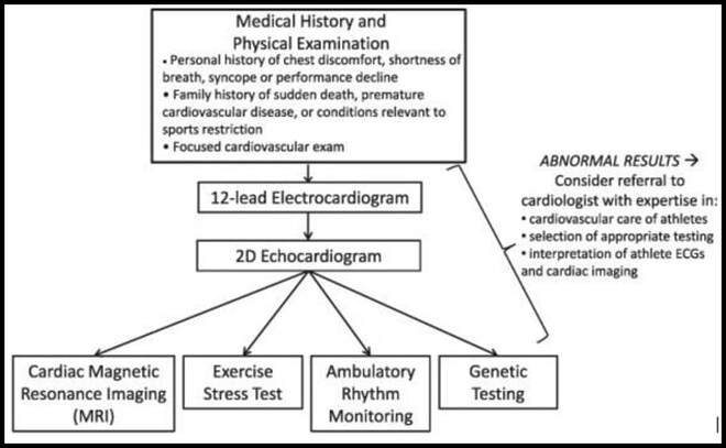
In contrast to the American recommendations, the European Society of Cardiology (ESC) and International Olympic Committee (IOC) advocate for screening that also includes a resting 12-lead electrocardiogram (ECG)
  • The role of the ECG in preparticipation screening has garnered considerable debate
  • ECG-inclusive screening appears to increase the sensitivity of preparticipation screening for identifying cardiovascular disorders that predispose to SCD
  • ECG in athlete screening remains unclear, and athletic programs should consider using ECG-inclusive screening based on the characteristics of their athlete population, the local screening resources available, and access to expert ECG interpretation specific to athletes.
 
 
Anatomic/Physiologic Changes 
  • Sports with significant isometric exercise may induce left ventricular hypertrophy to the same range as that of mild HCM (“gray zone” hypertrophy)
  • A small but significant proportion of endurance athletes will have dilated left ventricular (LV) cavities with low normal LV function, which overlaps with findings of a dilated cardiomyopathy
    • These physiologic changes to the left ventricle may be accompanied by right ventricular (RV) dilation and reduced systolic function, which could raise concern for ARVC in the appropriate context
 
 
SUMMARY:
Although rare, SCD in the athlete is a traumatic event that has a large impact on society
 
Incidence of SCD varies widely depending on the athlete population
  • Older athletes, SCD is primarily due to CAD and associated complications
  • Younger athletes, it is due to congenital or genetically mediated cardiovascular disease, such as HCM, coronary artery anomalies, other cardiomyopathies, or primary arrhythmogenic disorders
 
All preparticipation screening programs aimed at identifying athletes at high risk of SCD begin with a focused history and physical.
 
The addition of the 12-lead ECG and/or additional cardiac testing is a source of considerable ongoing debate.
 
It is highly unlikely that any screening program will be effective at appropriately identifying all athletes at risk of SCD; therefore, increased access to automated external defibrillators as well as training in cardiopulmonary resuscitation at a community level are important means of reducing SCD in athletes.
0 Comments

11/19/2018 0 Comments

Exertion Heat-related Illness


  • Results in more than 600 deaths a year in the United States!
  • Anticholinergic agents, beta-blockers, and sympathomimetic drugs can all interfere with heat removal and increase risk of heat-related illnesses
  • Patients with exertion related heat stroke are commonly young, athletes, or military personnel that present with symptoms after strenuous exercise in the heat
 
Heat Exhaustion
  • Occurs via water depletion or sodium depletion or combination
  • Water depletion occurs in elderly and persons working in hot environments
  • Salt depletion occurs when fluid losses are replaced with hypotonic solutions
 
  • Signs & Symptoms
    • Known heat exposure with temperature >37 C with:
      • Tachycardia/Palpitations
      • N/V
      • Diaphoresis
      • HA/malaise/fatigue/generalized weakness
      • Lightheadedness
      • Mentation is normal* (key distinguishing factor from heat stroke)
 
  • Evaluation
    • Labs may show:
      • Hemoconcentration
      • Entire spectrum of sodium derangements common electrolyte abnormality
 
  • Treatment
    • *NEED to remove from heat-stressed environment
    • Volume and electrolyte replacement as needed
    • Oral fluids vs. IVF 
    • Aggressive cooling (see below)
 
Heat Stroke
  • Severe end of heat-related illness spectrum with loss of thermoregulatory mechanisms
  • *TRUE EMERGENCY - focus of management should be on immediate, rapid cooling (even if can be started in pre-hospital setting!)
  • Mortality = 21-63%; can approach 30% even with treatment
  • Hallmark = Elevated temperature >41°C (106°F) + MSOF; heat exhaustion CAN have temperatures >104F
  • Occurs when endogenous heat production in combination with absorbed ambient heat exceeds the ability of the body to dissipate heat through adaptive mechanisms (i.e. sweating, hyperventilating, peripheral vasodilation)
  • The extent of neurologic injury and mortality is directly related to the peak temperature and duration of the hyperthermia

  • Symptoms
    • CNS is particularly susceptible -> AMS, coma, ataxia, confusion, seizures, hallucinations
    • Anhidrosis is frequently present; however, sweating found in up to 50% of patients
    • Shunting of perfusion to less vital organs (e.g. liver, gut) -> GI bleeds, ischemic hepatopathy
    • Compartment syndrome
    • *Hepatic injury is so common (↑AST/ALT) that if not present, consider an alternative diagnosis
  • Workup
    • ECG
    • Continuous core temp monitoring 
    • Blood glucose, CBC, CMP (including liver enzymes)
    • VBG (with lactic acid)
    • DIC labs – fibrinogen, D-dimer, PT/INR
    • CK (Rhabdomyolysis – 5x ULN) and UA (myoglobinuria)
    • Chest x-ray
    • CT brain (± LP), if indicated – BE SURE THIS IS NOT MENINGITIS
  • Management
    • As always in emergency medicine -> ABCs
    • Remove from environment
    • IVF (for renal protection and avoiding rhabdomyolysis)
      1. Goal UOP 3 mL/kg/hr
      2. Accumulation of intracellular cytoplasmic calcium which leads to myocyte cell membrane damage and ATP depletion -> Hyperphosphatemia and hypocalcemia
      3. Precise guidelines do not exist, but the goal CK should be a level less than 1000 U/L
      4. No target value for creatinine level, however may see ATN
    • Rapid cooling is mainstay of treatment
      1. Reduces morbidity/mortality, needs to be started in prehospital setting
        • Weak evidence available for target temperature; theoretical concern of overshoot hypothermia
      2. No role for antipyretics!
    • Techniques:
      1. ***Cool water immersion
        • Immersion of body to level of torso or neck in cool or ice-water
        • May not be immediately available, so consider other techniques (listed below)
        • As you can imagine, not well-tolerated
        • You will have to take them out of the water in the unfortunate event a patient has a cardiac arrest subsequently
      2. Diffuse application of ice or cold packs to entire body 
        • Benefit may be similar to ice-water immersion
      3. Evaporative/Convective Cooling
        • Set-up large fans and spray tepid water on patient’s body
        • Can perform this while getting ice bath drawn
        • Slightly higher morbidity and mortality compared to immersion
      4. Invasive
        • V V-ECMO, cold water rectal/bladder/NG lavage; bilateral chest tubes with pleural lavage… Weak data, so perform only if needed with caution
  • Consider giving antibiotics in addition to IVF hydration as it is difficult to rule out infection as a predisposing factor to the development of heatstrok
0 Comments

10/26/2018 0 Comments

LisFranc Fracture


Introduction
  • Disruption between the articulation of the medial cuneiform and base of the second metatarsal
    • Disruption of the TMT joint complex
    • Injury to 2nd metatarsal àDisruption of other metatarsals
  • May take form of purely ligamentous injuries or fracture-dislocations
    • Ligamentous vs. bony injury pattern has treatment implications
  • More common in males in their 30s 
  • Missed injuries can result in progressive foot deformity, chronic pain and dysfunction
    • Tarsometatarsal fracture-dislocations are easily missed (up to 30% on initial visits) and diagnosis is critical

Mechanism & Pathoanatomy
  • Mechanism = indirect rotational forces and axial load through hyperplantar flexed forefoot 
    • Hyperflexion/compression/abduction moment exerted on forefoot and transmitted to the TMT articulation
    • Metatarsals displaced in dorsal/lateral direction 
    • Think of a running back trying to push through lineman or sprinter
 
Anatomy
  • Midfoot:
    • Medial, Intermediate (Middle), Lateral Cuneiform
    • Navicular
    • Cuboid
  • Lisfranc joint complex consists of three articulations:
    • 1) Tarsometatarsal articulation
    • 2) Intermetatarsal articulation
    • 3) Intertarsal articulations
  • Ligaments
    • Lisfranc ligament  
      • Stabilizes 2nd metatarsal and maintenance of the midfoot arch
      • Interosseous ligament that goes from medial cuneiform to base of 2nd metatarsal on plantar surface 
    • No direct ligamentous attachment between first and second metatarsal

Physical Exam
  • Symptoms
    • Severe pain and inability to bear weight
  • PE
    • Medial plantar bruising with swelling throughout midfoot
    • Tenderness over tarsometatarsal joint
    • Pain with pronation and passive abduction of the midfoot
    • *Instability test
      • Grasp metatarsal heads and apply dorsal force to forefoot while other hand palpates the TMT joints
      • Dorsal subluxation suggests instability
      • If first and second metatarsals can be displaced, global instability is present, and surgery is required
      • When plantar ligaments are intact, dorsal subluxation does not occur with stress exam and injury may be treated nonoperatively
    • Evaluate for compartment syndrome!
 
Imaging
  • Radiographs
    • AP, lateral, oblique views
    • Stress -> may be helpful to show instability when non-weight bearing radiographs are normal and there is high suspicion
    • Weight-bearing with comparison view
    • 5 critical radiographic signsthat indicate presence of midfoot instability
      • 1) Discontinuity of a line drawn from the medial base of the 2nd metatarsal to the medial side of the middle cuneiform (diagnostic)
      • 2) Widening of the interval between the 1st and 2nd metatarsal
        • Fleck sign= bony fragment in 1st intermetatarsal space àAvulsion of Lisfranc ligament from base of 2nd metatarsal (diagnostic)
      • 3) Dorsal displacement of the proximal base of the 1st or 2nd metatarsal  
      • 4) Medial side of the base of the 4th metatarsal does not line up with medial side of cuboid 
      • 5) Disruption of the medial column line (line tangential to the medial aspect of the navicular and the medial cuneiform)
  • CT scan
    • Useful for diagnosis and preoperative planning
  • MRI
    • Can be used to confirm presence of purely ligamentous injury

Treatment
  • Nonoperative
    • Posterior splint and non-weightbearing for sprains and non-displaced fractures
      • No displacement on weight-bearing and stress radiographs and no evidence of bony injury on CT (usually dorsal sprains)
    • Cast immobilization for 8 weeks 
  • Operative
    • ORIF
    • Any evidence of instability (> 2mm shift)     
    • Favored in bony fracture dislocations as opposed to purely ligamentous injuries
    • Primary arthrodesis 
      • Purely ligamentous arch injuries   
      • Primary arthodesis is an alternative to ORIF in patients with any evidence of instability with possible benefits 
0 Comments

10/25/2018 0 Comments

Patellar Dislocation


Background
  • Typically occurs with trauma to an extended knee with externally rotated foot (quick pivoting in basketball, football and soccer)
  • Acute dislocation occurs with traumatic injury, M=F, may see hemarthrosis
  • Chronic dislocation or patholaxity seen more commonly in women
    • Ehlers-Danlos syndrome
    • Typically, little or no swelling; painless
    • Recurrent subluxation episodes with each flexion movement
  • May occur from a direct blow (ex. helmet to knee collision in football; knee-knee collision in basketball)
    • Usually on noncontact twisting injury with the knee extended and foot externally rotated
    • Patient will usually reflexively contract quadriceps thereby reducing the patella
  • Common associated fractures
    • Medial patella facet
    • Lateral femoral condyle
 
Clinical Features
  • Patella is usually displaced laterally
  • Knee is held in flexion
  • Acute dislocation usually associated with a large hemarthrosis   
    • Absence of swelling supports ligamentous laxity and chronic dislocation mechanism
 
Evaluation
  • Clinical diagnosis
  • May consider pre-reduction x-ray if concern for fracture (not required)
 
Management
  • Reduction = Relocation with lateral pressure on dislocated patella
  • Do NOT need x-rays prior to reduction
    • Consider XR following reduction to rule out fracture or loose body
  • Rarely need any sedation (sub-dissociative dose ketamine ?)
  • Option #1:
    • Mild flexion of hip (20-30 degrees by raising head of bed, not by propping the leg up off the bed) to relax quadriceps
    • Slowly extend and slightly hyperextend the knee and slide patella back into place.
  • Option #2
    • One provider applies slow downward pressure over the quads. This stretches out the muscle and slowly straightens the leg
    • At the same time, second provider pulls gentle traction of the patella outward while rotating the patella back over from lateral to anterior

​Disposition

  • Obtain ortho consult if unable to reduce or fracture/loose bodies seen on post-reduction x-ray
  • Otherwise may be discharged with ortho follow-up in 1-2 weeks
  • Non-Operative:
    • NSAIDS, activity modification, and physical therapy
    • Knee immobilizer for comfort (short-term)
    • Quad strengthening exercises
    • Core and hip strengthening to improve limb positioning and balance (hip abductors, gluteals, and abdominals) 
  • Consider knee aspiration for tense effusion
    • Positive fat globules indicates fracture
0 Comments

9/21/2018 0 Comments

Knee Dislocations


Introduction
  • Usually from a high-impact injury (tackle while leg planted); 
  • Outside of sports medicine, we typically see in trauma setting with dashboard injuries
  • Nearly 75% of ligaments will be disrupted 
  • Associated Injuries:
    • Vascular
      • 5-15% in all dislocation with nearly have occurring in A/P dislocations
    • Nerve
      • Common peroneal nerve injury in 25% of cases!
        • S: Decreased sensation over dorsum of foot sparing the lateral most portion
        • M: loss of dorsiflexion/eversion of the ankle; Loss of EHL -> Foot drop
    • Fractures presenting 60% of cases with femur and tibia being most common 
 
Signs & Symptoms
  • Symptoms = Knee pain/instability (brought to knees)
  • PE:
    • No deformity!
      • 50% of knee dislocations will spontaneously reduce prior to arrival; very deceptive
    • Swelling, effusion, abrasions
    • Obvious deformity
      • Reduce immediately!!!
  • Make sure you assess all ligaments (ACL, PCL, MCL, LCL)
  • Vascular exam is key!
    • Number one priority
    • Need to do both BEFORE and AFTER reduction
    • DP/PT pulses 
      • If pulses present/normal àDoes NOT rule-out arterial injury; collateral circulation can mask complete popliteal artery occlusion
      • ABI time!
        • >0.9 = serial examinations
        • <0.9 = CT angiography; consult vascular surgery if needed
      • Diminished/Absent Pulses:
        • Make sure knee is reduced!
        • Time = Muscle
        • IMMEDIATE surgical consult if pulses are still absent following reduction or hard signs of vascular compromise (expanding hematoma, distal ischemia, thrill)
        • Ischemia >8 hours has amputation rate ~85%
    • Pulses present after reduction àABIs àObservation vs angiography
 
Classification
  • Kennedy classification for direction of displacement of tibia on femur; Schenck for ligaments
  • Anterior
    • Most common (30-50%)
    • Hyperextension injury and usually requires PCL to tear
  • Posterior
    • 2ndmost common (25%)
    • Due to axial load on a flex knee
    • *Highest rate of vascular injury, i.e. complete popliteal artery tear
  • Lateral and Medical
    • Due to varus/valgus force
    • Lateral dislocation has highest change of peroneal nerve injury
 
Diagnostics:
  • XR ànormal if spontaneous reduction
  • Look for other fractures (Segond)
  • Non-emergent MRI
  • CT angiography as above


Treatment:
  • Reduce and re-exam!
  • This is an ORTHOPEDIC/SPORTS EMERGENCY
  • There is one situation where reduction of the knee should not take place.  The “dimple sign” represents buttonholing of the medial femoral condyle through the anterior medial joint capsule.  The sign indicates an irreducible dislocation and closed reduction is contraindicated for the risk of skin necrosis.
  • Posterior:
    • Someone holds distal femur, provider pulls longitudinally then anteriorly
    • Be careful; too much force can cause popliteal injury
  • Anterior:
    • Counter-traction on proximal tibia
  • Once reduced, splint in 20-30 (deg) flexion and re-shoot films
  • Most cases require surgical stabilization, consult your orthopedic surgeons
  • If ABI >0.9 with strong pulses and normal doppler -> Admit for observation
  • ABI <0.9 with asymmetric pulses or abnormal doppler -> consult vascular surgery, obtain CTA
0 Comments

9/14/2018 0 Comments

Clavicle Fracture


Background
  • Secondary to shoulder trauma (not as much direct clavicle trauma)
    • Tackled to ground
    • FOOSH
    • Direct impact to lateral shoulder
  • Majority of fractures involves the middle third (75-80%); Distal third (15%); Medial third (5%)
  • Distal third associated with AC joint injury and/or coracoclavicular ligament rupture
  • Medial fracture àintrathoracic injuries
 
Clinical Features
  • Swelling, deformity, tenderness, pain over anterior shoulder
  • Patient may be supporting affected arm with the contralateral arm
  • Displaced fractures
    • Medial fragment displaced posteriorly and superiorly (SCM pulls)
    • Lateral fragment displaced inferiorly and medically (Pectoralis and weight of arm pulls)
    • Open fractures usually result from medial fragment buttonholing through platysma
 
Evaluation
  • Assess distal pulses, motor, and sensation
  • XR (CXR, SXR, Dedicated clavicle films)
    • Associated injuries are rare, but can see rib fractures, PTX, neurovascular injuries
    • Additional XR view = ZANCA view (15-degree cephalic tilt)
      • Determines superior/inferior displacement
      • Have patient hold 5-10 lbs weight in affected hand
  • Consider CT for further eval and possible vascular injury
 
Classification
  • Neer, Allman, Craig, Robinson
  • AO Classification:
    • Type A, B, C (A = Simple; B = Wedge; C = Complex)
 
Management
  • Non-operative
    • Sling or figure-of-eight immobilization with early ROM exercises at 2-4 weeks; start strength training at 6-10 weeks
    • Best for minimally displaced, <2 cm shortening, no neuro deficits
    • Nonunion rate = 1-5 %
      • Most often in elderly and females
  • Operative
    • ORIF
      • Open fractures
      • Displaced fracture with skin tenting
      • Neurovascular injury
      • Risk of need for future procedures, implant removal or debridement for infection
  • Pain control
    • Chang AK, Bijur PE, Esses D, et al. Effect of a Single Dose of Oral Opioid and Nonopioid Analgesics on Acute Extremity Pain in the Emergency Department: A Randomized Clinical Trial. JAMA2017;318:1661-7
 
Disposition
  • Discharge with ortho/sports follow-up
  • Possible surgical intervention for comminuted fractures, significant displacement or >2 cm of shortening
  • Consult orthopedics in the ED if patient has an open fracture, Neurovascular compromise or skin tenting
0 Comments

9/14/2018 0 Comments

BiCeps Tendon Rupture


Incidence
  • Rare
  • Distal biceps tendon rupture represents about 10% of biceps ruptures, with majority being proximal
  • Ruptures tend to occur in the dominant elbow (86%) of men (93%) in their 40s
Risk factors
  • Your muscle builders with their anabolic steroids
  • Smoking has 7.5x greater risk than nonsmokers
  • Typically, patients will report a snap or pop as the elbow is eccentrically loaded from flexion to extension

  • Weakness and pain, primarily in supination, are hallmarks of this injury
  • May produce mid-arm "ball", also be referred to as the reverse “Popeye’s sign” = change in contour of the muscle
  • Motor exam often shows loss of supination and flexion, with mostly loss of supination
  • Distal swelling and tenderness over antecubital fossa
    • For most complete tears, there will be an Inability to palpate distal biceps tendon in antecubital fossa
    • However, there is a great provocative test you can perform called the Hook test. This was a technique first described in 2007 in an article published in the AJSM (Dr. Shawn O’Driscoll), which was found to be ~100% sensitive and specific
      • Performed by asking the patient to actively flex the elbow to 90° and to fully supinate the forearm. Using you index finger, attempt to hook the lateral edge of the biceps tendon.
      • With an intact / partially torn tendon, finger can be inserted 1 cm beneath the tendon
    • Follow-up study using the Hook test has shown once again near 100& specificity, however lower sensitivity at 82%, but overall a great physical exam maneuver if you have high suspicion!
  • The biggest challenge is going to be distinguishing between complete tear and partial tears
  • Biceps tendon is absent in complete rupture and palpable in partial rupture (otherwise they have a very similar clinical picture)

Evaluation
  • Obtain radiographs to rule-out avulsion fracture (Usually normal)
    •  However, occasionally shows a small fleck or avulsion of bone from the radial tuberosity
  • Ultrasound can help with diagnosis--> Use linear probe (high frequency probe) to assess for tendon defects
  • Ideally, outpatient MRI will often be used to distinguish between complete tear vs. partial tear or muscle substance vs. tendon tear
Management
  • Proximal rupture
    • Sling, ice, NSAIDS, physical therapy, referral to ortho
  • Distal
    • Nonoperative
      • Supportive treatment followed by physical therapy
      • Older, low-demand or sedentary patients who are willing to sacrifice function. Most becoming asymptomatic at 4-6 weeks
    • Operative
      • Surgical repair of by fixation of tendon to tuberosity 
      • Indications
        • Young healthy patients who do not want to sacrifice function 
        • Partial tears that do not respond to nonoperative management
        • Surgical treatment should occur within a few weeks from the date of injury
        • Further delay may preclude a straightforward, primary repair
0 Comments

9/7/2018 0 Comments

Achilles Tendon Injury


Background:
  • Associated with fluoroquinolone use
  • Sudden, severe pain typically with rapid acceleration or pivoting. Sports like football, track and field (sprinting), basketball, soccer
  • May hear a "pop"
  • Inability to run, stand on toes, or climb stairs
  • Most frequently ruptures 2-6cm above calcaneus (where blood supply is weakest)
 
Dx:
  • Clinical diagnosis
  • Ultrasound can be used in equivocal cases:
    • Use linear probe (high frequency probe)
    • Place probe in longitudinal plane over suspect tendon:
    • Achilles 2-6cm above calcaneus
    • Fan and slide side to side to optimize your view
    • Slide distal to proximal to find defect
    • Turn probe 90° to assess for tendon body defects
    • Positive Findings
      • Discontinuity in longitudinal view of ligament
      • Collection of fluid in longitudinal or transverse view suggests injury
    • Look at other limb for "normal" anatomy
    • Have patient range attempt plantar/dorsi-flexion and view in real time
    • Be aware of partial tears!
  • Comparing to normal ankle can reveal smaller defects or tears
    • Kankle
  • Thompson test (SN 96% and SP 93%)
    • Lay patient prone with knee bent at 90°
    • In normal patient, squeezing calf results in plantar-flexion; Patient’s with tendon rupture will likely have lack of plantar flexion with calf squeeze
  • Palpable defect in Achilles tendon 2-6 cm proximal to calcaneus, however the data on this finding isn’t very strong and wouldn’t base your diagnosis solely on this fact
 
Tx:
  • Short leg posterior splint in plantarflexion
  • ~25% of ruptures will have some amount of active plantar flexion or be able to walk
  • Even if partial tear, splint! Can evolve into a complete tear
  • Outpatient orthopedic referral 
 
0 Comments

9/7/2018 0 Comments

Mallet FInger Injury


​Background:
  • Usually caused by a traumatic impaction blow (i.e. sudden forced flexion) to the tip of the finger in the extended position. Often seen in ball sport like basketball, volleyball, and baseball. Commonly, I’ve seen this mostly in basketball players who present after “jamming” their finger when going for a rebound. Essentially, this blow forces the DIP joint into forced flexion, which the DIP doesn’t like. So in response, you get a rupture of extensor tendon in area of distal phalanx distal to DIP joint
 
PE:
  • DIP joint flexed to ~45°, lack of active DIP extension
 
Dx:
  • Clinical diagnosis
  • Consider finger x-ray (PA and lateral) to evaluate for avulsion fracture
  • Now I’m sure you all know about Doyle’s classification of mallet finger injuries? Uh, yeah, of course. Yeah, me either. Let’s leave that to the hand surgeons. 
 
Tx:
  • Now, most importantly… If left untreated, a mallet finger can lead to a swan neck deformity (PIP extension with DIP flexion; FDP unopposed)
  • For ED treatment, Splint DIP joint in continuous slight hyperextension, but don’t overextend x 6-8 weeks
  • Splinting of the PIP joint is not necessary and should be avoided; Let that PIP flow free!
    • Inadvertently splinting PIP for 6 weeks results in collateral ligamentous overgrowth and functional disability
  • Discharge with hand surgery follow-up in 7-10 days
 
 
Aluminum and Foam strips
Advantages:
  • Light weight and readily available
Disadvantages:
  • Must be taped in place
  • Patient cannot readily wash their hands while wearing
  • The foam will degrade over time
  • Skin can get macerated from lack of air getting to the skin
  • Splint limits finger use
 
Stax Splint
Advantages:
  • Lightweight and holds the fingertip straight
 Disadvantages:
  • Must be taped on
  • Covers the fingertip so patient’s finger cannot be used
  • Difficult to wash hands while wearing
  • Skin gets macerated
  • Can be difficult to fit
 
Oval-8® Finger Splint
Advantages:
  • One or two Oval-8 Finger Splints may be used to hold the DIP joint in extension while still allowing flexion of the proximal interphalangeal (PIP) joint
  • Lightweight-- the fingertip is open for function
  • Open design eliminates skin maceration
  • Size adjustability designed into the Oval-8 accommodates changes in swelling
  • Patient can wash hands easily by removing the splint
  • Easy to slide on and off for skin care
Disadvantages:
  • Professional adjustments may be needed for a custom fit
  • May fall off if weather is cold or swelling decreases
 
0 Comments

    Archives

    May 2019
    November 2018
    October 2018
    September 2018

    Categories

    All Sports Medicine

    RSS Feed

EMGuideWire

Picture
From the J. Lee Garvey Innovation Studio in the 
Department of Emergency Medicine
Carolinas Medical Center
Charlotte, NC

Picture
© COPYRIGHT 2015. ALL RIGHTS RESERVED.
LEGAL DISCLAIMER (to make sure that we are all clear about this):The information on this website and podcasts are the opinions of the authors solely.

For Health Care Practitioners: This website and its associated products are provided only for medical education purposes. Although the editors have made every effort to provide the most up-to-date evidence-based medical information, this writing should not necessarily be considered the standard of care and may not reflect individual practices in other geographic locations.

​For the Public
: This website and its associated products are not intended to be a substitute for professional medical advice, diagnosis, or treatment. Your physician or other qualified health care provider should be contacted with any questions you may have regarding a medical condition. Do not disregard professional medical advice or delay seeking it based on information from this writing. Relying on information provided in this website and podcast is done at your own risk. In the event of a medical emergency, contact your physician or call 9-1-1 immediately.