EMERGENCY MEDICINE GUIDEWIRE
  • HOME
  • The Podcasts
    • Core Concepts
    • Critical Care Topics
    • Intern Nuggets
    • Pediatric EM
    • Toxicology
    • Trauma Topics
    • Sports Medicine Corner
  • Cards for EM
    • ECG Monthly Lesson
  • CMC Imaging Mastery Project
    • Adult Chest X-Ray Cases
    • Pediatric X-Ray Cases
    • Adult Orthopedic X-Ray Cases >
      • Sternal Fractures
      • Native Hip Dislocations
      • Pelvic Ring Injuries
      • Shoulder Dislocations
    • Pediatric Orthopedic X-Ray Cases
    • Abdominal Imaging Room
    • NeuroImaging >
      • Head CT Interpretation Primer
      • Atraumatic Intracranial Infections
      • Cerebral Venous Sinus Thrombosis
      • Acute Epidural Hematomas
      • Subdural Hematomas
      • Subarachnoid Hemorrhage
      • Nail Gun Injury
      • IVROBA
      • Neurocysticercosis
    • Implanted Device Radiology Room
    • Condition Specific Radiology >
      • Aortic Coarctation
      • Lung Cancer
      • Pleural Effusion
      • Pneumonia
      • Septic Pulmonary Emboli
      • Spontaneous Pneumothorax
      • Blunt Aortic Injury
      • Aortic Dissection
      • Acute Chest Syndrome
      • Pericardial Effusion
      • COVID-19
      • Diaphragm Injury
      • Pneumomediastinum
      • Hypertrophic Cardiomyopathy
      • Situs Anomalies
      • Stress-Induced Cardiomyopathy
      • Mechanical Circulatory Support Devices
      • Peripartum Cardiomyopathy
      • Sternal Fractures and Dislocations
      • VENTRICULOPERITONEAL SHUNTS
  • Global EM
  • Shownotes
    • Core Concepts ShowNotes
    • Cards for EM ShowNotes
    • Ped EM Morsels ReBaked Shownotes
    • Pediatric EM Shownotes
    • Sports Med Shownotes
    • Toxicology Shownotes
    • Trauma Talks Shownotes
  • EM Advanced Clinical Topics Sympsosia
    • EM:ACT Critical Care
    • EM:ACT Pediatric EM
    • EM:ACT Trauma
    • EM:ACT Cardiac Emergencies
    • EM:ACT Gastrointestinal Emergencies
    • EM:ACT Women's Health Emergencies
  • Subscribe
    • iTunes
    • Google Play
    • Stitcher
  • About
  • Associated Sites
    • CMCEdMasters
    • CMCECGMasters
    • PedEMMorsels
    • Dr. Sofiya Diurba - Checkpoint Inhibitors
    • Human Trafficking
Search by typing & pressing enter

YOUR CART

8/16/2018 0 Comments

Pain Management in Our ED


Sergey Motov, MD
@painfreeED
 
General Principles:
  1. Management of acute pain in the ED should be patient-centered and pan syndrome specific by using multimodal approach that include non-pharmacological modalities and pharmacological ones that include non-opioid and opioid analgesics.
  2. Assessment of acute pain should be based on a need for analgesics to improve functionality, rather than patients-reported pain scores.
  3. ED clinicians should engage patients in shared decision-making about overall treatment goals and expectations, the natural trajectory of the specific painful condition, and analgesic options including short-term and long-term benefits and risks of adverse effects.
  4. When opioids are used for acute pain, ED clinicians should combine them with non-pharmacologic and non-opioid pharmacologic therapy: Yoga, exercise, cognitive behavioral therapy, complementary/alternative medical therapies (acupuncture); NSAID’s, Acetaminophen, Topical Analgesics, Nerve blocks, etc.
  5. When considering opioids for acute pain, ED clinicians should involve patients in shared decision-making about analgesic options and opioid alternatives, risks and benefits of opioid therapies, and rational expectations about the pain trajectory and management approach.
  6. When consider opioids for acute pain, EM providers should counsel patients regarding serious adverse effects such as sedation and respiratory depression; pruritus and constipation, and rapid development of tolerance and hyperalgesia
  7. If acute pain lasting beyond the expected duration, complications of acute pain should be ruled out and transition to non-opioid therapy and non-pharmacological therapy should be attempted 
  8. When considering administration of opioids for acute pain, ED providers should make every effort to accesses respective state’s Prescription Drug Monitoring Program (PDMP). The data obtained from PDMP’s to be used to identify excessive dosages and dangerous combinations, identify and counsel patients with opioid use disorder, offer referral for addiction treatment.
0 Comments

8/9/2018 0 Comments

What's in your Code Cart? Know your Meds


0 Comments

8/5/2018 0 Comments

Hyperemesis Gravidarum


Hyperemesis Gravidarum:
  • Hyperemesis Gravidarum is severe nausea and vomiting during early pregnancy, typically starting prior to 9 weeks gestation.
    • NOT associated with significant pain
    • Do not turn off your brain, rule out serious causes before diagnosing the patient with hyperemesis gravidarum
  • Workup includes basic chemistry and urinalysis to evaluate electrolytes and signs of starvation ketosis
  • Confirm IUP
    • Molar pregnancies or multiple gestation pregnancies can cause a higher amount of HCG and these patients are more likely to have hyperemesis.

  • First line treatment for this condition is Vitamin B6 and doxylamine. Some women will respond to P6 acupressure or ginger pills.
  • Treatment includes fluids with a source of glucose and antiemetics.
    • Promethazine, metoclopromide, and ondansetron are all recommended options by the American College of Obstetrics and Gynecology (ACOG)[1].
    • Ondansetron
      • There are insufficient data on fetal safety with ondansetron use and further studies are warranted
      • There have been studies that fetal exposure to ondansetron increases the risk of pediatric cardiac abnormalities and cleft palate; however, the data is inconsistent.
      • Individual studies of the association between ondansetron and congenital malformations are inconsistent, with some showing an increase in birth defects and others showing no difference [2,3,4]. A recent systematic review of ondansetron use in early pregnancy found eight studies that were adequate for inclusion [5]. Although there was a small increase in the risk of cardiac defects in two of the studies (odds ratio [OR], 2.0; 95% CI; 1.3-3.1 and OR, 1.62; 95% CI; 1.04-2.14), there was no increase in the overall rate of malformations in the ondansetron-exposed patients.
      • ACOG says, “Although some studies have shown an increased risk of birth defects with early ondansetron use, other studies have not. The absolute risk to any fetus is low.”
      • The American College of Obstetrics and Gynecology recommend Ondansetron as an option for patients not controlled on the first line medications Vitamin B6 and doxylamine.
      • Consult with your local Obstetricians
 
  1. Erick, Miriam, et al. “ACOG Practice Bulletin 189.” Obstetrics & Gynecology, vol. 131, no. 5, 2018, p. 935., doi:10.1097/aog.0000000000002604.
  2. Pasternak B, Svanstrom H, Hviid A. Ondansetron in pregnancy and risk of adverse fetal outcomes [published erratum appears in N Engl J Med 2013;368:2146]. N Engl J Med 2013;368:814–23.
  3. Danielsson B, Wikner BN, Kallen B. Use of ondansetron during pregnancy and congenital malformations in the infant. Reprod Toxicol 2014;50:134–7.
  4. Einarson A, Maltepe C, Navioz Y, Kennedy D, Tan MP, Koren G. The safety of ondansetron for nausea and vomiting of pregnancy: a prospective comparative study. BJOG 2004;111:940–3.
  5. Carstairs SD. Ondansetron use in pregnancy and birth defects: a systematic review. Obstet Gynecol 2016;127:878–83.
 
0 Comments

8/2/2018 0 Comments

Pediatric Nasal Foreign Bodies


Background:
  • Most common site of foreign body insertion in children
  • Can be missed and remain for weeks or months
  • Population at risk:
    • Children between 2–6 years-old most common
    • Mental retardation
    • Psychiatric illness
  • Complications:
    • Sinusitis is the most common complication
    • Septal perforation
    • Bronchial aspiration
  • High risk of complications with button batteries:
    • Mucosa ischemia
    • Turbinate or septal damage -> Saddle-nose deformity
Etiology:
  • Organic vs. Inorganic
  • Button batteries: High risk of complications compared with other foreign bodies (tissue necrosis, septal perforation, saddle-nose deformity); require rapid removal!
    • Septal perforation can occur in as little as 4 hours
  • Magnets (Used to mimic nasal piercing)
    • May cause intestinal perforation if swallowed, especially newer high-powered neodymium magnets
Signs & Symptoms:
  • Most nasal foreign bodies are asymptomatic
  • Unilateral nasal obstruction with nasal pain
  • Purulent/foul-smelling nasal discharge
  • Persistent epistaxis
History:
  • Child witnessed putting object into nose or FB noticed by parent/caretaker
  • Many children are reluctant to admit to placing a foreign body for fear of adult disapproval
  • Child may present weeks after with nasal discharge and bleeding
  • *Often misdiagnosed at this stage as sinusitis
Diagnosis:
  • Laboratory studies usually unnecessary
  • Fiberoptic visualization if foreign body cannot be visualized on rhinoscopy
  • Sinus films if present for extended period or suspicion of battery or magnet
  • May need chest or abdomen films for aspiration/ingestion
Treatment:
  • Keep in sitting position to avoid posterior displacement and possible aspiration of foreign body
  • Avoid interventions that upset the child
  • Topical vasoconstrictors:
    • Nebulized epinephrine
    • Cocaine: 4%
    • Oxymetazoline: 0.05%
    • Phenylephrine: 0.125–0.5%
*NOT recommended for button batteries as may increase leakage of caustic materials
  • Hooked probe, alligator forceps
    • Used for anterior foreign bodies that are easily grasped
  • Headlamp, nasal speculum will facilitate use!
  • Suction catheter can be used for round, smooth objects
  • Balloon catheters (or Katz extractor) can be used primarily when instrumentation fails
    • Can use a 5F or 6F Foley or Fogarty balloon catheter lubricated with 2% lidocaine jelly
  • Positive pressure for children or the “parent’s kiss”
    • Positive pressure applied to mouth only 
    • Parent may tell the child he or she will be given a “big kiss.”
    • Deliver brisk puff as child begins to inhale
    • Alternatively, deliver puff with a bag-valve mask over the mouth and O2 at 10–15 L/min.
  • Procedural sedation may be necessary
    • Need availability of advanced airway as foreign body has potential for dislodgment posteriorly and aspiration!
  • Snare technique (for you MacGyver’s out there):
    • 24-gauge wire made into a loop with a hemostat
    • Slip through swollen tissue, behind object, pulling it free
Disposition:
  • Admission Criteria:
    • Foreign body cannot be recovered in ED or posteriorly displaced
    • Removal under general anesthesia is required
    • Mucosal ischemia or turbinate/septal damage
  • Discharge Criteria
    • Ensure that there is no airway compromise
    • If a button battery was removed, monitor for delayed sequelae as outpatient
    • May require follow-up with ENT if removal unsuccessful in ED or concern for nasal mucosa injury
 
 
0 Comments

7/14/2018 0 Comments

Upper GI Bleeds


Stable UGIB

DDx:
  1. PUD: most common cause. Associated with NSAID use and H.Pylori, smoking. 
  2. Erosive gastritis/esophagitis: Associated with alcohol, salicylates/NSAIDs, infectious, stress from severe illness (cushings)
  3. Varices: Portal HTN 
  4. Mallory- Weiss Syndrome: Repetitive vomiting
  5. Dieulafoy Lesion:
    1. Arteries that protrude through the GI submucosa and can cause intermittent bleeding with negative endoscopic findings. 
    2. NSAIDs and liver disease can predispose to this
  6. AVMs
  7. Malignancy
  8. Aortoenteric fisula
  9. Ischemia/perforation
  10. Epistaxis/Upper airway bleed
  11. Ingestions
Hx:
  1. Is this really a GI bleed? 
    1. Nosebleed
    2. Ingestions (guiac card) 
  1. Timing/amount
  2. H/o GIB
  1. If so h/o scope
  1. EtoH, NSAID, Smoking hx
  2. Anti-platlet /anti-coagulation history
  3. Good ROS
PE: 
  1. Vital signs
  2. Abdominal Exam
  3. Rectal Exam (guiaic)
  4. S/s Liver failure
    1. Jaundice
    2. Ascites
    3. Palmar erythema
    4. Spider nevi
    5. Caput medusa
    6. Hepatomegaly/splenomegaly
Labs:
  1. Hb/hct
  2. CBC
  3. Basic Chemistry
    1. BUN: Cr ?30 suggestive of bleed
  1. Coags
  2. Type and screen


Treatment:

  1.  PPIs 
    1. oral (omeprazole, pantoprazole, lansoprazole) vs IV therapy. No difference
    2. Continuous vs intermittent IV PPI boluses
      1. JAMA systematic review that showed 13 different RCTs revealing no difference between gtt vs bolus in mortality, rebleeding, LOS, PRBC transfusion  
    3. PPI vs placebo
      1. No difference in mortality
      2. Reduced surgical intervention
      3. Reduced Rebleeding (NNT = 15)
  1. Somatostatin (Octreotide-long acting somatostatin): 
    1. ​Decrease gastric acid, decrease blood flow to stomach/duodenum, splanchnic vasoconstriction
    2. 50 mcg bolus
  2. Antibiotics
    1. Cirrhotic patients are innately immunocompromised -> increased risk of gut bacterial translocation
    2. 1 g Rocephin
    3. ONLY Tx that has mortality benefit with NNT = 4 to prevent an infection NNT = 22 to save a life!
  3. Pro-motility agents
    1. Ertyhromycin/Azithromycin (500mg)
    2. Decreases need for second EGD
  4. Transfusion     
    1. Threshold use to be Hbg < 9, yet theres mortality benefit with transfusion of Hbg < 7
    2. If thrombocytopenia <50k with active GIB, transfuse platelets 

Disposition:

Risk scoring systems to help risk stratify

  1. Glasgow Blatchford Scale: 
    1. NPV of a GBS score of <1 for the outcome of transfusion, death or need for intervention was 99% 
    2. Approximately 20% of patients’ presenting with upper GI hemorrhage have a Blatchford score of zero
  1. Rockall Score
  2. AIM65 Score

Unstable UGIB

  1. Airway/breathing
    1. Preoxygenate
      1. Limit BVM
      2. Hbg dissociation curve shift right in anemia
    2. Upright
    3. RSI vs DSI
  2. Circulation
    1. MTP 1:1:1 Resuscitation
    2. Reverse Coagulopathy
      1. Cirrhotics —> Vit K IV, FFP, ? 4 factor FFP
      2. NOAC reversal: praxibind, kcentra
      3. Thrombocytopenia —> Plts
      4. ESRD/ASA —> DDAVP 0.3mcg/kg
    3. Permissive Hypotension —> MAP goal 60-65
    4. TXA —> HALT-IT data to come in May 2019.
  3. Medications
    1. PPI
    2. Octreotide
    3. Antibiotics
    4. Pressor of choice? Vasopressin
  4. Treatment/Consultants
    1. Ballon Tamponade 
      1. Call for xray
      2. 3 ports: Esophagus balloon, Gastric ballon, Suction
      3. Measure out your E and G with NGT at 50 cm mark on Blakemore 
      4. Insert it with lube
      5. Insufflate 50 ccs of air into gastric lavage port
      6. Insufflate 50 ccs of air into gastric port
      7. Xray, make sure ballon bubble below diaphragm
      8. Put another 200 ccs of air in gastric port for total 250 ccs
      9. Place on traction
      10. Insert your NGT to esophageal port
      11. Blakemore holds approximately 50-100ccs of air 
      12. Get manometer
      13. Goal manomatry for esophagus 30-45 mmHg
    2. Endoscopy
    3. IR for embodied vs emergent TIPs (Transjugular Intrahepatic Portosystemic Shunts)
    4. Surgery

Core Concepts:

  • Keep your DDx broad for UGIB and perform a focused H&P and PE
  • Use risk stratification tools like GBS, Rockall, and AIMS 65 to help with your disposition
  • Remember that the most important intervention that affects mortality is antibiotics in your cirrhotic UGIBs as well as restrictive transfusion with only those with Hbg < 7
  • Other treatment remedies include PPIs (no difference between IV vs oral or gtt vs bolus), somatostatin (or somatostatin analogs like octreotide), and promotility agents 
  • In a hemodynamically unstable UGIB, remember your ABCs. Secure your airway early, resuscitate with blood products, consider reversal of coagulopathies. 
  • Know how to place your hail mary Blakemore while making early consultations to your ICU, GI, IR and surgical colleagues.
0 Comments

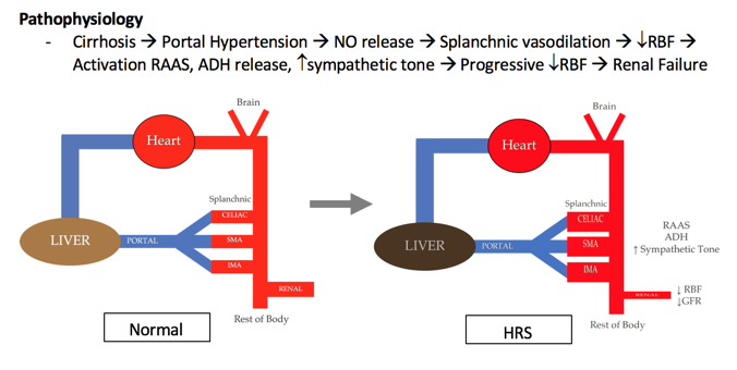
7/3/2018 0 Comments

Hepatorenal Syndrome

Hepatorenal Syndrome: Core Concepts
 
Hepatorenal Syndrome (HRS) = Liver Disease + Acute Kidney Injury (AKI)
  • Liver disease with portal hypertension with an AKI without other etiology
  • Diagnosis of exclusion
 
  • Type 1 HRS: Doubling of serum creatinine to Cr > 2.5mg/dL in less than 2 weeks
    • Median survival = 2 weeks
 
  • Type 2 HRS: Renal impairment less severe than HRS Type 1
    • Median survival = 6 months​
Picture
Treatment
  • Liver transplant is the only treatment that will reverse the disease pathology
  • Dialysis is used only a bridge while awaiting transplant
    • If patient is not a liver transplant candidate, dialysis is not offered as it does not treat the underlying pathology
  • TIPS procedure is only indicated if there is another acute process going on, like a GI bleed. Not indicated for HRS alone
 
Management in the ED
  • Identify patients with screening labs and urinalysis
  • Resuscitate with albumin (Goal 1g/kg over the first 24 hours)
  • Resuscitate further with vasoactive agents with goal of raising MAPS 10-15mmHg
    • Norepinephrine, vasopressin, and somatostatin can all be used for vasoconstriction, specifically at the splanchnic circulation
    • Look for inciting conditions such as infection 
0 Comments

5/6/2018 0 Comments

Acute Urinary Retention

Acute Urinary Retention Slides by Dr. Covell below:

0 Comments

4/12/2018 0 Comments

The Dying Asthma Patient


Outline The Dying Asthmatic Patient:
  • Critical asthma syndrome:
    • Inability to speak
    • PEFR< 25% predicted
    • Failed frequent bronchodilator/IV steroids
  • Start with the basics:
    • Inhaled bronchodilators: Beta agonists, anticholinergics
      • MDI versus Nebulizer: equally effective in mild/moderate asthma
      • Continuous nebs benefit in severe disease
    • Systemic beta-agonists
      • IM Terbutaline: 0.25 mg subq q20min
      • IM Epinephrine: 0.3-0.5 mg subq q20min
    • Steroids
      • Give within 1 hour to significantly decrease admission rates
      • Use oral prednisone/dexamethasone or in more severe cases IV methylprednisolone
    • IV Magnesium
      • 2 grams over 15 minutes to avoid hypotension
      • Significant improvement in pulmonary function in severe asthma exacerbations
  • Consider other therapies:
    • Heliox
      • Helium oxygen mixture 80:20
    • Ketamine:
      • Bronchodilation and vagolytic
  • Non-invasive pressure ventilation
    • IPAP=ventilation (high CO2=need IPAP)
    • EPAP=oxygenation (opens up alveoli)
    • Consider starting settings at IPAP 10 to EPAP 2 with FiO2 of 100%
  • Endotracheal intubation
    • Don’t do it too early, don’t do it too late
    • Consider DSI vs RSI
    • Permissive hypercapnia
  • Post intubation hypotension
    • SH!T: stacking, hypovolemia, induction drugs, tension pneumothorax
  • Last ditch efforts
    • Inhaled paralytics
    • VV ECMO
  • Core Concepts:
    • Start with the basics: nebs, steroids, mag
    • Remember the your back pocket tricks: heliox, ketamine, NIPPV
    • Intubation, don’t do it too early, don’t do it too late. Consider DSI vs RSI
    • Post intubation remember to manage your vents carefully including your i to e ration, avoiding barotrauma
    • With your post intubation hypotension mnemonic oh sh!t!: stacking, hypovolemia, induction drugs, tension pneumothorax
    • Know what hail marys are available at your hospital including inhaled anesthetics or ECMO
0 Comments

3/25/2018 0 Comments

INcreased ICP

Increased Intracranial Pressure
Russell Trigonis PGY-2, Katie Lupez PGY-2, and Chris Gardner PGY-1
 
  • Increased intracranial pressures (ICP) can have many different causes with traumatic brain injuries (TBI) being one of the most common ones we see in the ED
  • Intracranial pressure, mean arterial pressure (MAP), and cerebral perfusion pressure (CPP) are linked according to following formula
CPP = MAP - ICP
 
  • In situations where intracranial pressure is elevated by a factor like a traumatic bleed, cerebral perfusion pressure will if the MAP remains constant. If the MAP drops too, the CPP can decrease to a critical level at which point the brain becomes ischemic
  • Increased intracranial pressure can be suspected with changes in GCS, localizing symptoms, or hemodynamic changes like bradycardia and hypertension (Cushings reflex)
  • If you suspect increased ICP, evaluate them quickly focusing on the ABCDs with a good neuro exam
  • If GCS < 8 or appears to be declining quickly, intubate to protect the airway
 
  • Consider ketamine and rocuronium for intubation
    • Ketamine has been proven safe in patients with increased ICP and shows no additional increase in ICP when used for RSI followed by mechanical ventilation
    • Rocuronium has been associated with improved mortality compared to succinylcholine in TBI patients
  • Make first attempt best attempt for intubation. Use video laryngoscopy if available as your first line agent to allow proper immobilization of C-spine as well as better first pass success.
 
  • Passive techniques to allow maximal venous and CSF drainage to minimize ICP
    • Elevated head-of-bed or reverse Trendelenburg positioning (30 degrees)
    • Properly fitted and positioned c-collar
    • Low mean airway pressures
 
  • Active techniques to decrease ICP
    • Consider sedation and paralytics only if patient requires it (cough, agitation). Otherwise these agents can cause hypotension or minimize your ability to perform serial neurological exams]
    • Hypertonic saline (3% NaCl) with a bolus of 150 – 250cc in adults may be more beneficial than mannitol (1-3 g/kg) to avoid AKI and hypovolemia
    • Can also consider 2 Amps of Sodium Bicarbonate (100cc of ~6% hypertonic solution)
0 Comments

3/11/2018 0 Comments

MEthylene Blue use in the ED


​Methylene Blue Summary 
  • Uses include methemeglobinemia and vasoplegia. There is also some evidence supporting its use in angioedema.  
  • Dose for methemeglobinemia is 1.5mg/kg over 1-2min 
  • Dose for vasoplegia is 1-2mg/kg over 1-2min, then option infusion of 1-2mg/kg/hr over a maximum of 1-2hrs. 
  • Mechanism of action: lots of biochemistry involved, but it breaks down to methylene blue inhibiting soluble cGMP to prevent NO endothelium-dependent relaxation in vascular smooth muscle to halt NO induced vasodilation.  
  • Adverse effects of methylene blue administration include pain/burning at the injection site, nausea, HA 
  • Avoid use in patients with G6PD deficiency, and use with caution in patients on multiple serotonergic agents (SSRIs, SNRIs, TCAs) as methylene blue can induce serotonin syndrome. This last point is especially important when using the drug as an infusion as there is high risk for serotonin syndrome with prolonged use.  
  • Don’t forget to warn you nursing/support staff that the pulse ox reading may be artificially low while the methylene blue is being infused, however this reaction is transient.  
0 Comments
<<Previous
Forward>>

    Archives

    November 2020
    September 2020
    February 2020
    January 2020
    August 2019
    July 2019
    May 2019
    March 2019
    February 2019
    January 2019
    December 2018
    November 2018
    October 2018
    September 2018
    August 2018
    July 2018
    May 2018
    April 2018
    March 2018
    February 2018

    Categories

    All Core Concepts Pediatric

    RSS Feed

EMGuideWire

Picture
From the J. Lee Garvey Innovation Studio in the 
Department of Emergency Medicine
Carolinas Medical Center
Charlotte, NC

Picture
© COPYRIGHT 2015. ALL RIGHTS RESERVED.
LEGAL DISCLAIMER (to make sure that we are all clear about this):The information on this website and podcasts are the opinions of the authors solely.

For Health Care Practitioners: This website and its associated products are provided only for medical education purposes. Although the editors have made every effort to provide the most up-to-date evidence-based medical information, this writing should not necessarily be considered the standard of care and may not reflect individual practices in other geographic locations.

​For the Public
: This website and its associated products are not intended to be a substitute for professional medical advice, diagnosis, or treatment. Your physician or other qualified health care provider should be contacted with any questions you may have regarding a medical condition. Do not disregard professional medical advice or delay seeking it based on information from this writing. Relying on information provided in this website and podcast is done at your own risk. In the event of a medical emergency, contact your physician or call 9-1-1 immediately.